

Breast Conservation Surgery
Wide Local Excision / Lumpectomy
Breast Cancer Surgery Options
Breast cancer surgery involves two main components:
Surgery to the breast
Surgery to the lymph nodes
With respect to surgery on the breast, there are two main types of breast cancer surgery:
Breast conservation surgery (lumpectomy or wide local excision)
Mastectomy
The type of surgery recommended depends on a range of individual factors. Importantly, the type of breast surgery does not determine whether chemotherapy is required. A common misconception is that mastectomy reduces the need for chemotherapy; however, chemotherapy decisions are guided by tumour biology rather than the type of surgery performed.
What Is Breast Conservation Surgery?
Breast conservation surgery, also known as a lumpectomy or wide local excision, involves removal of the breast cancer along with a small margin of surrounding healthy tissue, while preserving the remainder of the breast.
This approach is most commonly used for small, early-stage cancers, many of which are detected through BreastScreen. The breast usually appears similar following surgery, although it may be slightly smaller and will have a scar.
Most women who undergo breast conservation surgery will require radiotherapy to the remaining breast tissue to reduce the risk of cancer recurrence. Approximately 70–80% of women with breast cancer are suitable for breast-conserving surgery.
What Factors Influence the Ability to Have Breast Conservation Surgery?
Breast Size and Tumour Size
Removing more than 10% of breast volume can result in noticeable changes to breast shape. Oncoplastic techniques allow safe removal of 20–30% of breast tissue while maintaining breast shape.
Smaller breasts are more prone to shape changes and may require oncoplastic approaches. If a tumour occupies 30–40% or more of the breast, a mastectomy is often required.
In some cases, chemotherapy before surgery may be used to shrink the tumour, allowing breast conservation or oncoplastic surgery instead of mastectomy.
Number of Tumours
If multiple tumours are present within the breast (multifocal breast cancer), a mastectomy is usually required.
When more than two tumours are suspected, an MRI is commonly performed to determine whether there are truly multiple cancers or a single larger tumour that was not clearly seen on mammogram or ultrasound.
Genetic Mutations
Younger patients with a confirmed BRCA1 or BRCA2 gene mutation may benefit from mastectomy, even for small tumours.
This option is discussed with all patients under the age of 40 who have a strong family history of breast cancer or a confirmed genetic mutation.
Ability to Have Radiation Therapy
Breast conservation surgery involves removal of the tumour followed by radiation therapy.
If radiation therapy is not possible or is declined, breast conservation is not suitable, and a mastectomy is recommended. A lumpectomy without radiation increases the risk of cancer recurrence by 5–6 times over five years compared with those who receive radiation.
Why Is Breast Radiation Needed?
Breast radiotherapy (XRT) uses X-rays to treat the remaining breast tissue after breast conservation surgery.
Treatment typically lasts 3–6 weeks, delivered Monday to Friday at a radiation centre.
Breast conservation surgery removes the visible cancer, but microscopic cancer cells may remain in the surrounding breast tissue. Radiotherapy significantly reduces the risk of recurrence and provides cancer outcomes equivalent to mastectomy while preserving breast appearance.
Five-Year Recurrence Rates
Mastectomy: 2–5% (0.5–1% per year)
Breast conservation + radiation: 5–7.5% (1–1.5% per year)
Breast conservation alone: 20–30% (5–6% per year)
Why Might I Need Further Surgery to Obtain Clear Margins?
Breast conservation surgery aims to remove the cancer with a margin of healthy tissue while preserving breast appearance.
The pathology report assesses whether margins are:
Clear
Close
Involved
An involved margin means cancer cells are present at the edge of the specimen and usually requires further surgery, known as cavity re-excision.
Close margins occur in approximately 20% of breast conservation cases. Modern radiotherapy has made smaller margins acceptable in some cases. In general:
A margin >1 mm is acceptable for invasive cancer
A margin >2 mm is acceptable for DCIS
Each case is assessed individually, and Dr Green will discuss whether additional surgery is required.
Will Breast Conservation Surgery Definitely Avoid a Mastectomy?
Breast conservation surgery does not guarantee that a mastectomy will not be required later.
Occasionally, the cancer may be larger than initially seen on imaging, requiring additional surgery. In patients with smaller breasts, removal of additional tissue may significantly affect breast shape, leading to recommendation of mastectomy with or without reconstruction.
If this becomes necessary, Dr Green will explain all options thoroughly.
What Is Oncoplastic Breast Surgery?
Oncoplastic breast surgery is now considered standard of care in modern breast cancer treatment.
It combines cancer surgery with plastic surgery techniques to:
Remove cancer safely
Maintain or improve breast shape
Reduce long-term deformity
Traditional surgery often resulted in noticeable contour defects and scarring. Oncoplastic techniques aim to prevent these issues and improve body image and satisfaction following treatment.
When Should Oncoplastic Breast Surgery Be Used?
Oncoplastic surgery should be available to all patients and is standard practice in many parts of Australia.
Studies show that removing more than 7–10% of breast tissue can lead to deformity. Oncoplastic techniques reduce this risk, even when smaller volumes are removed.
Historically, fluid cavities left after tumour removal could collapse after radiation, causing deformity later — often incorrectly attributed to radiation rather than surgical technique. Oncoplastic surgery addresses this issue.
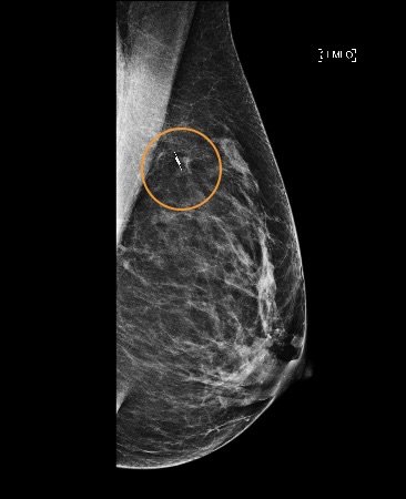
How Are Small Cancers Located During Surgery?
When a breast cancer cannot be felt, localisation techniques are required.
Traditionally, a hookwire is used. Dr Green now uses a radar reflector (SCOUT® locator) at St Andrew’s War Memorial Hospital.
The SCOUT reflector:
Is about the size of a grain of rice
Is placed using imaging prior to surgery
Allows real-time tumour localisation during surgery
Is accurate to within 1 mm
This precision allows exact tumour removal with minimal surrounding tissue, reducing deformity and the need for further surgery.
What Happens After My Operation?
Most patients stay in hospital for one night after breast conservation surgery.
Pain is usually minimal. You will be advised to wear a supportive bra day and night until your follow-up appointment. Detailed bra information is provided prior to surgery.
You will return approximately one week after surgery to:
Review pathology results
Discuss further treatment if required
Have dressings removed
Possible Complications of Breast Conservation Surgery
Breast conservation surgery may include a sentinel lymph node biopsy or axillary surgery. Please refer to those sections for further details.
Most patients recover well and are discharged within 1–2 days. However, complications can occur.
More Common (up to 10%)
Reduction in breast size
Bruising
Need for second operation to obtain clear margins (≈15%)
Long-term pain or discomfort
Uncommon (up to 5%)
Wound infection
Bleeding requiring re-operation
Poor cosmetic outcome
Keloid (raised) scarring
Need for third operation to obtain clear margins
Lymphoedema of the breast
Rare but Important (less than 1%)
Anaesthetic complications
Deep vein thrombosis or pulmonary embolism
Heart attack
Allergic reactions
Nipple necrosis
Severe breast deformity
This list is a guide and not exhaustive. Please discuss any concerns with Dr Green during your consultation.


Oncoplastic Breast Conservation
Therapeutic breast reduction ; removal of the breast cancer using a breast reduction technique.


Hookwire localisation


SCOUT Radar Localisation